We are a multi-disciplinary research group focused on developing quantitative biomarkers for non-invasive, real-time assessments of tissue structure and function to diagnose disease or trauma and guide therapies.
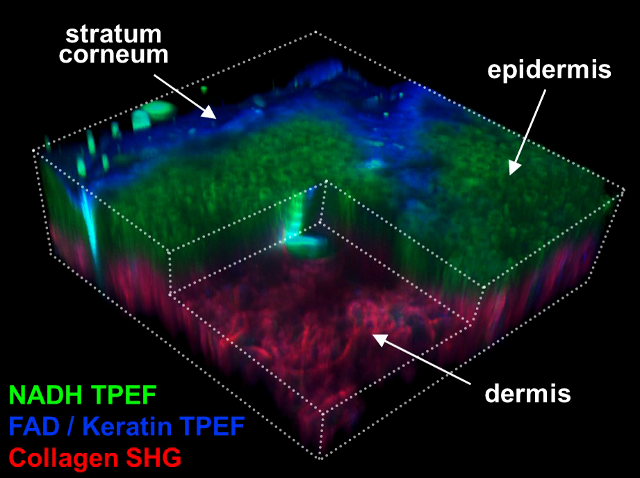
Label-free multiphoton microscopy can provide a variety of non-invasive quantitative biomarkers of cell metabolism, collagen fiber organization, and tissue composition. By quantifying the naturally present fluorescence of NADH and FAD without our cells, we can non-invasively measure dynamic changes in the metabolism of live cells, tissues, and organisms. Second harmonic generation (SHG) imaging can also provide a non-destructive measure of 3D collagen organization within tissues. While acquiring two-photon excited fluorescence (TPEF) and SHG signals, we can also measure coherent anti-Stokes Raman scattering (CARS), which provides images of a tissue’s protein and lipid composition. We combine these imaging methods with deep learning and advanced image analysis approaches to quantify the orientation, morphology, and fractal organization of different cell and tissue features.
The following selected press releases provide an overview of our techniques and how we are using them to solve various biomedical research and health-related problems:
Our research primarily focuses on skin wound healing and aging applications in which we can integrate label-free multiphoton microscopy with advanced image analysis and mechanical testing. However, we are always interested in collaborating with researchers in other areas where non-invasive multiphoton microscopy or advanced image analysis of tissue microstructure can aid in scientific discovery and advancements in medicine. Below are a list of our active research collaborations that have produced publications or secured significant funding:
- Dr. Aristidis Veves, Beth Israel Deaconess Medical Center, Harvard Medical School
- NIH R01AG056560, In vivo label-free characterization of aged skin to predict delayed wound healing (PI: Quinn)
- Quinn KP, Leal EC, Tellechea A, Kafanas A, Auster ME, Veves A, Georgakoudi I (2016). Diabetic wounds exhibit distinct microstructural and metabolic heterogeneity through label-free multiphoton microscopy. Journal of Investigative Dermatology, 136(1) 342-4.
- Dr. Ed Sander, University of Iowa
- NIH R01AG056560, In vivo label-free characterization of aged skin to predict delayed wound healing (PI: Quinn)
- Dr. Francois Berthiaume, Rutgers University
- DoD W81XWH-17-1-0194, Nanoparticle systems for pressure wound healing (PI: Berthiaume)
- Dr. Jeff Wolchok, University of Arkansas- Biomedical Engineering
- Roberts K, Schluns J, Walker A, Jones JD, Quinn KP, Hestekin J, Wolchok JC (2017). Cell derived extracellular matrix fibers synthesized using sacrificial hollow fiber membranes. Biomedical Materials, 13 (1), 015023.
- Dr. Jingyi Chen & Dr. Suresh Kumar, University of Arkansas- Chemistry & Biochemistry
- Chancellor’s Discovery, Creativity, Innovation, and Collaboration Fund, Monitoring the delivery of hyper-stable growth factors to skin wounds through label-free multiphoton microscopy (PI: Quinn)
- Dr. Kartik Balachandran, University of Arkansas- Biomedical Engineering
- Lam NT, Muldoon TJ, Quinn KP, Rajaram N, Balachandran K (2016). Valve interstitial cell contractile strength and metabolic state are dependent on its shape. Integrative Biology, 8 (10), 1079-1089.
- Dr. Martin Yarmush, MGH, Harvard Medical School & Dr. Alex Golberg, Tel Aviv University
- Li X, Saeidi N, Villiger M, Albadawi H, Jones JD, Quinn KP, Austen WG, Golberg A, Yarmush ML (2018). Rejuvenation of aged rat skin with pulsed electric fields. Journal of Tissue Engineering and Regenerative Medicine, 12, 2309-2318.
- Golberg A, Villiger M, Khan S, Quinn KP, Bouma BE, Mihm MC, Austen WG, Yarmush ML (2016). Preventing scars after injury with partial irreversible electroporation. Journal of Investigative Dermatology, 136 (11), 2297-2304.
- Dr. Narasimhan Rajaram, University of Arkansas- Biomedical Engineering
- Lam NT, Muldoon TJ, Quinn KP, Rajaram N, Balachandran K (2016). Valve interstitial cell contractile strength and metabolic state are dependent on its shape. Integrative Biology, 8 (10), 1079-1089.
- Alhallak K, Rebello LG, Muldoon TJ, Quinn KP, Rajaram N (2016). Optical redox ratio identifies metastatic postential-dependent changes in breast cancer cell metabolism. Biomedical Optics Express, 7 (11), 4364-4374.
- Alhallak K, Jenkins S, Lee DE, Greene NP, Quinn KP, Griffin RJ, Dings RPM, Rajaram N. Optical imaging of radiation-induced metabolic changes in radiation-sensitive and resistant cancer cells (2017). Journal of Biomedical Optics, 22 (6), 060502-060502.
- Lee D, Alhallak K, Jenkins SV, Vargas I, Greene NP, Quinn KP, Griffin RJ, Dings RPM, Rajaram N (2018). A Radiosensitizing Inhibitor of HIF-1 alters the Optical Redox State of Human Lung Cancer Cells In Vitro. Scientific Reports, 8(1), 8815.
- Vargas I, Alhallak K, Kolenc OI, Jenkins SV, Griffin RJ, Dings PRM, Rajaram N, Quinn KP (2018). Rapid quantification of mitochondrial fractal dimension in individual cells. Biomedical Optics Express, 9(11), 5269-79.
- Dr. Michele Pierro, Vivonics Inc.
- DoD W81XWH-17-C-0169, Non-contact Tissue Viability Assessment (PI: Pierro)
- Dr. Shilpa Iyer, University of Arkansas- Biological Sciences
- NIH R21HD094394, Development of quantitative biomarkers for mitochondrial disorders (Multi-PI: Quinn & Iyer)
- Dr. Tim Muldoon, University of Arkansas- Biomedical Engineering
- Lam NT, Muldoon TJ, Quinn KP, Rajaram N, Balachandran K (2016). Valve interstitial cell contractile strength and metabolic state are dependent on its shape. Integrative Biology, 8 (10), 1079-1089.
- Alhallak K, Rebello LG, Muldoon TJ, Quinn KP, Rajaram N (2016). Optical redox ratio identifies metastatic postential-dependent changes in breast cancer cell metabolism. Biomedical Optics Express, 7 (11), 4364-4374.
For more information, please contact Dr. Kyle Quinn (kpquinn AT uark.edu).